
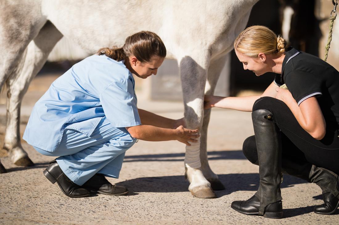
Lameness is a general term that refers to a horse with an abnormal gait or stance. This is a common issue that requires assessment by a veterinarian to determine the cause and best course of action.
Common causes of lameness include strain or injury, acute or chronic laminitis, genetic traits, infection, metabolic issues, or neural disorders.
A timely lameness exam can identify the underlying cause and how to manage it to relieve pain and support longevity.
A lameness exam typically involves discussing the horse’s history, observing the horse at rest and in motion, performing flexion tests and using hoof testers to identify sources of pain. Further diagnostic tests may be needed if these assessments are not sufficient.
Lameness evaluations are also a crucial part of pre-purchase exams. Identifying possible lameness in a horse you are interested in purchasing allows you to determine if you would like to proceed with the purchase. If you purchase the horse, lameness exams can indicate what may become an issue in the future.

Types of Lameness
According to the American College of Veterinary Surgeons, lameness is defined as an abnormality of a horse’s gait or stance. Lameness is not a disease itself but is a clinical sign that can be caused by pain, restrained movement or neuromuscular dysfunction.
Lameness is usually caused by pain in the muscles, tendons, bones, ligaments, or joints. Less commonly, non-painful lameness can also occur from neurological dysfunction. [1]
Most lameness can be separated into the following categories:
- Front end lameness
- Hind end lameness
- Pain in the back and sacroiliac
- Compensatory lameness
- Neurological lameness
Front End Lameness
Front leg lameness is the easiest to see with even the untrained eye. Front end lameness produces the classic “limp” one would expect to see with a painful limb.
When looking to see if a horse is lame in front, look for two features of the gait:
- Head bobbing
- Differences in range of motion
When stepping on a painful limb, most horses will use the placement of their head to try to alleviate pressure on that limb. Horses will raise their head when they step on a sore limb to reduce pressure on that limb and lower it when they step with the non-painful limb.
When trotting your horse on a circle or straight line, pay attention to when they lift and lower their head. If you are only able to trot your horse on a circle, make sure you are looking at their gait in both directions, as they could be lame on both limbs. [3]
If a horse has a sore muscle or painful joint, it will likely exhibit a shortened stride length and reduced range of motion, referring to how far a limb can move with little pain in a gait cycle and how much flexion they can tolerate without a pain response. [4]
Horses should move their limbs evenly and smoothly during a gait cycle. Any hitching or unevenness can indicate pain. [3]
When a horse is lame in both front legs, for example with laminitis, it is more difficult to detect because there is no head bob. Instead, look for:
- Rigid head carriage – either high or low
- Loss of the rhythmical swing to the back at the walk
- Bracing of the back
- Tension in the forearms, shoulders, back and hindquarters
- Reluctance to walk or make sharp turns
- Greatly shortened stride length
Hind End Lameness
Hind end lameness can be subtle, with most horse owners noticing that something is “off” without being able to put their finger on the source. Horses lame in behind may exhibit:
- An uneven rise and fall of the hips and buttock
- Differences in range of motion
Horses that are lame in behind will drop the hip on the sore side slightly more than the non-painful side and may produce odd-looking movement patterns in their hind limb joints to compensate for painful areas.
For example, twisting in hock motion is common with pain in this area, and horses with stifle pain will often point the foot outward and rotate the stifle outward.
Like front-end lameness, horses will often show differences in their range of motion when lame in the hind legs. The lame leg may not come forward underneath the horse’s body or extend out as far behind the body as much as the non-painful leg. [3]
Back and Sacroiliac Pain
Back and sacroiliac pain can cause movement abnormalities and behavioural issues. Your horse may have a painful back and sacroiliac region if they exhibit: [5]
- Discomfort during grooming or pressure on the back
- Abnormally short strides
- Resistance to saddling or girthiness
- Behavioural issues during performance such as refusing fences, bucking, rearing, etc.
Compensatory Lameness
When a horse experiences pain or discomfort in one area of the body they often develop compensatory movements to alleviate pain in the affected area. For example, if the horse is lame in one forelimb it may alter its gait and weight-bearing to shift more weight onto the other forelimb.
Unfortunately, this can lead to compensatory lameness in the previously healthy limb. Compensatory lameness can be difficult to distinguish from the original source of lameness.
Asymmetry in the vertical movement of the withers can help distinguish primary and compensatory lameness when both front limbs are affected. [14]
Neurological Lameness
Lameness due to a neurological disease can be subtle, extremely obvious or somewhere in between. It may begin in early life or following an infection.
Early in the disease progression, your horse may exhibit:
- Subtle abnormalities in their range of motion, which come and go
- Minor stumbling or tripping
- Issues retaining balance when turning in small circles
- Reluctance when transitioning from one ground surface to another
- Weakness and inability to balance when a leg is held up
Late in the disease progression, your horse may exhibit: [6]
- A wide stance when standing or walking
- Swaying back and forth
- Low muscle tone in the tail and anus
- Frequent falling
- Seizures
- Inability to step over ledges or other low obstacles
- Loss of balance if you raise their head
- Muscle atrophy
Lameness due to a neurological disease or injury is always very serious and needs a thorough work-up by your vet.
Examples of neurological diseases that can cause incoordination include Wobbler syndrome, equine protozoal myeloencephalitis (EPM) and equine herpesvirus type 1 myeloencephalopathy.
Botulism also causes ataxia, weakness, tongue and anal tone weakness in the early stages before progressing to inability to rise and respiratory paralysis. Rabies must also always be on the list of possibilities.
Nutritional deficits can also lead to poor coordination and lameness. For example, Vitamin E deficiency can cause a serious condition called equine motor neuron disease with ataxia, weakness and muscle mass loss. [17]
Genetic conditions can also cause incoordination and lameness. One example is neuroaxonal dystrophy involving the brainstem found in Morgan horses. Signs of these typically appear within the first 2 years of life and have a variable prognosis. When the condition also involves the spinal cord it is called equine degenerative myeloencephalopathy. There is evidence that vitamin E status impacts the severity. [18]
Lameness Exams
When performing a lameness exam, your veterinarian will follow a protocol that maximizes the likelihood of identifying the location of pain while minimizing the need for major diagnostic tests.
The American Association of Equine Practitioners uses the following scale to help owners and veterinarians identify the severity of lameness. On this scale, a grade of 0 means no lameness and 5 means extreme lameness.[2]
Lameness Scale
- Grade 0: Lameness is not perceptible under any circumstances; manipulating the limbs does not produce gait abnormalities.
- Grade 1: Lameness is difficult to observe and is not consistently apparent regardless of circumstances.
- Grade 2: Lameness is difficult to observe at a walk or trot in a straight line but consistently apparent in certain circumstances.
- Grade 3: Lameness is observable at a trot under all circumstances.
- Grade 4: Lameness is obvious at a walk.
- Grade 5: Lameness produces a complete inability to bear weight or a complete inability to move.
Medical History
The first thing your veterinarian will do is evaluate your horse’s medical history. They will want to know if your horse has any past lameness issues and what signs you have observed during the current episode.
This exam will include questions such as: [14]
- What type of exercise does your horse do? Did lameness first appear during exercise?
- How long has the horse been lame? Has it been stable or gotten worse/better?
- Have they been rested or exercised since lameness first appeared?
- Does lameness improve as they warm up to exercise?
- When is the lameness most consistently evident?
- Have you given any treatments? What were the effects?
- When was the horse last shod or trimmed? What is their typical farrier care?
- What abnormalities do you notice when riding or watching the horse?
Additional information such as your horse’s age, pedigree and past use can also be valuable to determine underlying contributing factors.
Prevalence
Depending on the type of work the horse does they may be more prone to certain lameness conditions. For example, racehorses are more prone to lameness associated with repetitive overuse such as foot bruising, fractures and suspensory injuries.
In comparison, draft horses are more likely to become lame due to hoof cracks and abscesses or osteoarthritis. [14]
Competitive and working Western horses are prone to hock and high suspensory injuries.
Knowing the type of work, intensity and training history will help your veterinarian understand your horse’s likelihood of certain injuries or conditions.
Observing Your Horse at Rest
Your veterinarian will want to study your horse while they are standing still on flat ground. They will evaluate your horse’s conformation, how they are weight-bearing at rest, and whether there are any obvious signs of strain. [14]
Observations will be made from a distance and up close. From farther away your vet can evaluate stance, frequency of weight shifting, unusual limb positioning, body conformation and body condition.
From up close, your vet can assess the feet for conformation and balance, hoof cracks, hoof size and abnormal wear. Joints and tendons can be inspected for pain, heat, and swelling, and muscles can be inspected for heat, pain, swelling and/or atrophy.
Your veterinarian will emphasize comparing one side of the body to the other to identify the source of lameness.
Observing Your Horse in Motion
As part of diagnosing lameness, your vet will want to see your horse walk and trot in a straight line. They will observe your horse from the front, back, and sides as they walk and jog in hand.
They may want to watch your horse walk, trot, and canter in a circle to see how they move in the gait and how they transition between gaits.
Your vet will look for any gait deviations such as plating or winging, altered range of motion and abnormal placement of the feet.
This will help identify abnormal movement asymmetries in the body that can indicate the source and severity of lameness. Slight asymmetries in head and pelvic movements are a normal part of biological variability between horses, but larger asymmetries could indicate an issue. [15]
Assessments should be done under several circumstances to avoid over-interpretation, including on soft and hard surfaces, in a straight line and while lungeing. This will help distinguish between normal asymmetries vs asymmetries that indicate lameness.
For example, normal asymmetries in head and pelvic movements can appear in sound horses when lungeing but not when trotting in a straight line. [15][16]
Asymmetries might also not be the same in both directions. These natural asymmetries could mimic or mask limb lameness. Therefore, several observations during various forms of exercise should be made.
Hands-on Exam
Your veterinarian will perform a thorough hands-on exam involving palpation of the hooves, limbs or other areas that may be sources of pain. They will pay attention to any areas of tenderness, swelling, heat, or other physical abnormalities that could have resulted from an injury.
This should include systematic palpation of the pastern, fetlock and metacarpus/metatarsus as well as the higher joints in the leg, plus the neck, back and sacroiliac region. Palpation of muscles should also be done to identify areas of scarring, spasm, swelling or weakness.
Hands-on exams are usually done following observations at rest, although some veterinarians may also do this assessment following exercise. [14]
Application of Hoof Testers
If your vet thinks your horse’s hooves are sources of pain, they will perform a hoof test. Hoof testers are a blunt, scissor-like tool that can be used to apply pressure to certain areas of the hoof. If your horse has painful hooves, they will react to the applied force by pulling back or taking the foot away.

Horses that show sensitivity over broad areas of the sole might have a fracture in the distal phalanx, extensive bruising or laminitis. If the horse is sensitive in small, specific areas this could indicate localized bruising, puncture wounds or abscesses. [14]
Horses with navicular area pain tend to react to pressure across the heels and over the frog. [21]
Hoof testers can also indicate hoof wall issues, such as chronic laminitis or white line disease.
However, it is not unusual for a horse with laminitis to not react to hoof testers. This may be because having to bear all the weight on the other laminitic foot is more painful. [22]
Flexion Tests
Flexion tests can help reveal lameness that may not otherwise be apparent. Your veterinarian will hold your horse’s leg flexed for a pre-determined period, anywhere from 5 to 60 seconds. [14]
They will then release the leg and ask you to immediately trot the horse in a straight line away from them. Your vet will watch for signs of pain, stiffness, improper weight shifting, or irregular movement.
This test can be used to identify severity of damage to an affected joint by assessing the horse’s response to light or firm flexion.
A scoring system should be used to track responses in various joints and to track improvement over time as your horse recovers.
Diagnostic Tests
If your veterinarian cannot pinpoint the exact cause and location of the lameness or if they think a major issue is causing the abnormal gait, they will likely suggest further diagnostic testing. These tests can offer a more precise explanation and help clarify which treatment would be most effective.
Nerve Blocking
A nerve block is used when it is difficult to pinpoint the exact location of the pain. A local anesthetic will be injected into specific areas of your horse’s body to render them numb.
These locations are often near joints or particular parts of the hoof. Once the anesthetic has taken effect, you will trot your horse away from and towards the veterinarian to determine whether the horse is now sound.
If your horse trots away sound, the vet knows that the blocked area was the source of pain. If they trot away lame, this area was not of the issue and other tests are needed. [7]
Local anesthetic injected directly into a joint will sometimes make the lameness worse. This is also considered a positive test and likely indicates the joint is inflamed.
Radiographs
If your veterinarian thinks that bone issues are the cause of lameness, they will suggest radiographs (x-rays). Most radiographs can be performed at the barn, but those covering large areas such as the back and sacroiliac may need to be performed at a veterinary clinic.
Radiographs provide limited information regarding soft tissue injuries (i.e., tendon, ligament, bursa). If the issue involves soft tissue, your vet may need to perform an ultrasound. [8]
Ultrasound
When performing an ultrasound, your vet will apply a jelly-like substance to your horse’s skin. This gel provides a medium through which the ultrasound waves can travel.
The ultrasound wand transmits ultrasonic waves which bounce off your horse’s tissues, producing an image. Your vet will examine the image for soft tissue irregularities. [9]
Rectal ultrasounds can also be performed if upper limb lameness is suspected. This exam can identify fractured vertebrae, muscle inflammation, pelvic fractures, hip problems, sacroiliac issues or irregularities in blood vessels that may be contributing to lameness. [14]
MRI and CT Scans
Magnetic resonance imaging (MRI) and computed tomography (CT) are advanced imaging techniques that provide detail on both bone and soft tissue anatomy. They are used more often in small animals because of their size. The need for anesthesia to prevent movement artifacts also complicates their use in horses.
MRI scanning is more commonly used in horses than CT and is now available at most veterinary hospitals. [19] A portable machine for standing MRI is also available and in use by both universities and full service clinics. It can be used to image the fetlock or foot with the horse under standing sedation. [20]
Scintigraphy (Nuclear Scanning)
If the source of lameness remains unclear, your vet may suggest a nuclear scan. This procedure requires injecting your horse with a radioactive substance and it is always performed in a veterinary hospital.
After your horse arrives at the hospital, they will be injected with a radioactive isotope called Technitium. Technitium is taken up in areas with abnormal inflammation or metabolic activity.
Your horse is then scanned with a Gamma camera that picks up areas of Technitium absorption, indicating “hot spots” of injury and pain. [10]
Arthroscopy
If your veterinarian has pinpointed the source of lameness to a specific joint but does not know the exact cause, they may suggest an arthroscopy. Under general anesthesia, a small scope is inserted into your horse’s joint to visualize the interior joint structures.
If a cause for lameness is found, surgery to correct the issue is often performed at the same time. [11]
Synovial Fluid Sampling
If joint infection or inflammation is suspected, your vet may suggest taking a synovial fluid sample.
A small needle is inserted into the affected joint, and synovial fluid is aspirated and collected. This can be examined for markers indicting inflammation and infection. [12]
Neurological exams
Horses that show poor coordination or odd limb placement may have a neurological condition. A thorough neurological exam involves:
- watching the horse move
- reflex testing
- evaluating the response to putting limbs in abnormal locations
- cranial nerve testing
- skin sensation testing
- visual field testing
- ophthalmoscopic exam
- measuring vitamin E levels
- sampling the cerebrospinal fluid for cytology and titres
- genetic testing to identify known cause, if indicated
Radiographs or advanced imaging may be recommended if spinal or brain disease is suspected.
During a lameness exam, your vet may perform a tail pull test. A healthy horse will resist being pulled to one side by the tail. However, horses with neurological issues may appear weak and offer little resistance to being moved. [14]
Pre-Purchase Exams
One instance in which a lameness exam is warranted is during a pre-purchase exam (PPE). Buying a horse is an expensive undertaking, so many potential buyers will ask a vet to perform a PPE to highlight any issues the horse may have.
A PPE can determine if the horse is lame at the time of the exam. During a PPE, your vet will perform:
- A physical exam to identify past injuries and conformation issues
- Lameness exam in hand and under saddle
- Flexion tests
- Radiographs
- A blood sample to test for pain masking drugs such phenylbutazone or for tranquilizers
A prepurchase exam is NOT supposed to speculate on the horse’s likely future soundness, only on how the horse moved that day. However, radiographs may detect areas of abnormality that may cause problems in the future.
Veterinarians can tailor PPEs to the intended use of the horse. For example, a PPE for an upper-level dressage horse may look different from a PPE for a lower-level hunter. [13]
Preventing Lameness Issues
Many causes of lameness are avoidable with the right management program. You can reduce the likelihood of wear and tear on your horse by:
- Implementing appropriate feeding and exercise programs for growing horses
- Getting and maintaining a meticulously balanced trim that has the horse landing heel first and flat on all four hooves
- Not pushing your horse past what they are conditioned for
- Feeling your horse’s legs every day for any heat and swelling
- Icing or cold hosing your horse’s legs after particularly hard work sessions
- Warming your horse up appropriately before working them hard
- Avoiding work in muddy or deep footing
- Maintaining a healthy body condition
- Meeting their vitamin and mineral requirements
- Supporting your horse’s joints with scientifically validated supplements
Lameness can be complicated to diagnose and treat. Keep a vigilant watch for anything that may appear abnormal in your horse’s movements. Prompt diagnosis and treatment can save you money and keep your horse working well into their career.
If your horse is lame, work with an equine nutritionist to formulate a diet that supports joint health and recovery from lameness. You can submit your horse’s information online for a free evaluation.
References
- American College of Veterinary Surgeons. Lameness in Horses. Large Animal Topics. 2022
- American Association of Equine Practitioners. AAEP Lameness Scale. 2005
- Keegan, K. Head Movement Pattern in Horses With Forelimb and Hindlimb Lameness. AAEP Proc. 2005.
- Lea, R and Gerhardt, J. Range-of-motion measurements. The Journal of Bone and Joint Surgery. 1995.
- Allen, K et al. How to Diagnose and Treat Back Pain in the Horse. AAEP Proc. 2010.
- Tyler, C et al. A survey of neurological diseases in horses. Aus Vet Journal. 1993.
- Drevemo, S et al. Nerve block and intra-articular anaesthesia of the forelimb In the sound horse. Equine Vet Journal. 2010.
- Auer, J and Fackelman G. Treatment of Degenerative Joint Disease of the Horse: A Review and Commentary. Vet Surg. 1981.
- Reef, V. The Use of Diagnostic Ultrasound in the Horse. Ultrasound Quarterly. 1991.
- Archer, D et al. Skeletal scintigraphy in the horse: Current indications and validity as a diagnostic test. The Vet Journal. 2007.
- Muttini, A et al. Arthroscopy in the Horse. Vet Research Comm. 2003.
- Tulamo, R et al. Sequential clinical and synovial fluid changes associated with acute infectious arthritis in the horse. Equine Vet Journal. 1989.
- Burba, D. Pre-purchase Examination. Man of Clin Proc in the Horse. 2017.
- Baxter, G.M. et al. Examination for Lameness: History, Visual Exam, And Conformation. Lameness in Horses 7th Ed. 2020.
- Hardeman, A.M. et al.Variation in gait parameters used for objective lameness assessment in sound horses at the trot on the straight line and the lunge. Equine Vet J. 2019.
- Rhodin, M. et al. Head and pelvic movement asymmetry during lungeing in horses with symmetrical movement on the straight. Equine Vet J. 2016.
- Mohammed, H.O. et al. Vitamin E deficiency and risk of equine motor neuron disease. Acta Vet Scand. 2007.
- Finno, C.J. and Johnson, A.L. Equine Neuroaxonal Dystrophy and Degenerative Myeloencephalopathy. Vet Clin North Am Equine Pract. 2022.
- Magnetic Resonance Imaging (MRI). PennVet. Accessed April 8, 2024.
- Giorio, M.E. et al. Presence and size of synovial masses within the navicular bursa correlate well between magnetic resonance imaging and bursoscopy and have a guarded prognosis. Equine Vet J. 2023.
- Sardari, K. et al. Differentiation of Heel Pain from Other Hoof Pain in Horses. JAppl Anim Res. 2005.
- Rietmann, T.R. et al. The Association between Heart Rate, Heart Rate Variability, Endocrine andBehavioural Pain Measures in Horses Suffering from Laminitis. J Vet Med. 2004.


Leave A Comment